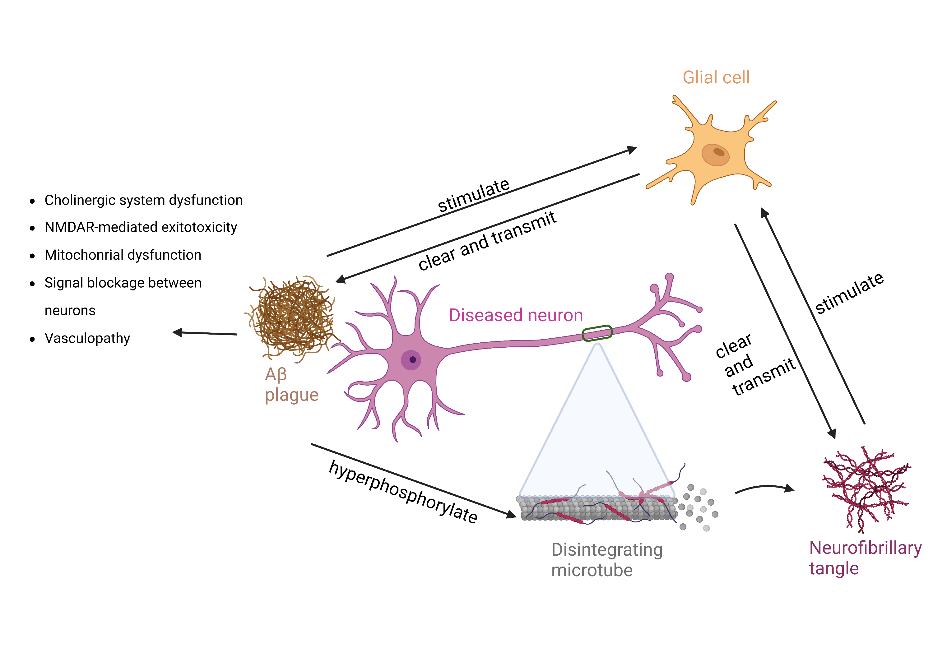
Alzheimers disease (AD) is a common neurodegenerative disease. The histopathological changes of AD include amyloid β-protein (Aβ) deposition, tau tangles, neuroinflammation and neurodegeneration. Some of the pathological changes could be shown in vivo by positron emission tomography (PET) and magnetic resonance imaging (MRI) biomarkers which play a key role in diagnosing AD. Fluorodeoxyglucose positron emission tomography (FDG-PET) can reflect and predict dysfunction. Aβ-PET was sensitive for the diagnosis of early AD but cannot distinguish the severity of AD. Tau-PET can compensate for the deficiency of Aβ-PET. Tau tangles were positively correlated with the severity of AD, and also associated with cognitive impairment. Probes targeting neuroinflammation of AD have been developed, but further study is needed to validate its effectiveness. Conventional MRI performs high tissue contrast that can show structural changes and has been routinely applied in clinical practice, such as evaluation of cerebral atrophy. Advanced MRI sequences (such as DTI、ASL、MRS、BOLD and QSM) that can provide additional information beyond structure that includes brain microstructure, blood perfusion, metabolite concentration, brain activity, connections and networks between brain regions, iron deposition, etc. The integrated PET and MR may improve the diagnostic efficiency of AD.